Zebrafish could help scientists fully restore human vision loss
Discover how zebrafish retinal regeneration offers insights into reversing vision loss through restored photoreceptor functionality and neural integration.
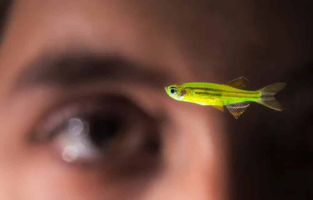
Zebrafish’s ability to regenerate retinal cells provides hope for restoring human vision. Learn how cutting-edge research is uncovering potential therapies. (CREDIT: Keith Walters ’11)
Neuronal degeneration leading to visual impairment or blindness remains a critical challenge in human health. Unlike humans, zebrafish possess a remarkable ability to regenerate retinal neurons after injury. This capacity provides a unique opportunity to explore potential therapies for human vision loss.
Zebrafish exhibit this regeneration through their Müller glia (MG) cells, which act as stem cells. Upon retinal damage, MG cells re-enter the cell cycle, producing neuronal progenitors that proliferate and differentiate into retinal cells. This process restores the retina’s cellular and synaptic layers. While structural regeneration is evident, functional recovery of regenerated retinal neurons remains a significant hurdle.
Two fundamental questions arise when studying retinal regeneration: Do regenerated neurons recover normal light-evoked responses, and can they integrate into existing circuits? To address this, researchers developed a novel approach to monitor neural activity in zebrafish cone photoreceptor terminals.
Using a combination of intense UV light lesions and advanced imaging techniques, scientists induced retinal damage and tracked the functional recovery of photoreceptors.
UV light-induced lesions ablate photoreceptor cells in a wavelength-dependent manner, with complete histological recovery achieved within 28 days. Researchers found that regenerated UV cones exhibited light-evoked responses comparable to those in an undamaged retina. Over three months, these cones restored both Off- and On-signals—a testament to their reintegration into the horizontal cell (HC) network.
"The presence of both response types highlights the recovery of intrinsic photoreceptor function and their connectivity with neighboring cells," explained a lead researcher. This discovery underscores the potential of zebrafish as a model for studying retinal regeneration and functional restoration.
Assessing functional recovery required groundbreaking tools. Zebrafish cones regenerate with restored chromatic responses and functional connectivity. Researchers leveraged a customized two-photon imaging setup combined with calcium imaging and light stimulation. This approach allowed them to track photoreceptor activity at synapses, where signals are passed to neighboring nerve cells.
A significant challenge was overcoming the interference of light stimulation with microscopy observations. Collaborations between institutions led to the development of a bespoke microscope, capable of isolating observation from stimulation.
Related Stories
This innovation enabled scientists to examine light responses at various wavelengths, revealing that regenerated photoreceptors transmit signals with the same sensitivity, quality, and speed as intact ones.
Prof. Michael Brand, leader of the study, emphasized, "Our results confirm that regenerated photoreceptors not only regain their physiological functions but also reintegrate into neural circuits."
Unlike mammals, zebrafish regenerate photoreceptors through MG cells. In humans, MG cells lack this regenerative capacity due to evolutionary changes. However, their structural similarities suggest the potential for therapeutic applications.
“Mammalian MG cells resemble those of zebrafish. This similarity offers hope that their regenerative potential could be reactivated,” stated Prof. Brand. The team’s findings provide a framework for exploring human retinal repair, aiming to develop therapies for conditions like retinitis pigmentosa and macular degeneration.
Current human therapies involve either stimulating retinal stem cells or transplanting lab-grown photoreceptors. However, functional integration of these cells remains a key challenge. By studying zebrafish, researchers can identify mechanisms to enhance connectivity and signal transmission in human treatments.
Vision restoration hinges on recreating the intricate interactions between photoreceptors and neural circuits. The zebrafish model has proven that regeneration from endogenous stem cells can restore vision at a functional level. While zebrafish have evolved to regenerate seamlessly, humans might need biotechnological interventions to achieve similar outcomes.
The research team’s findings, published in Developmental Cell, mark a significant step toward understanding the regenerative process. Using zebrafish, they demonstrated that regenerated photoreceptors regain full functionality. They respond to light stimuli, transmit signals, and integrate into existing networks effectively.
“Our study shows the potential for human applications. While this is basic research, it lays the groundwork for revolutionary treatments,” Prof. Brand explained. With advancements in genetic engineering and molecular biology, researchers hope to mimic zebrafish regeneration in humans, potentially transforming the treatment landscape for blinding diseases.
Human vision relies on the retina, a neural tissue that captures light and converts it into electrical signals. Damage to photoreceptor cells results in irreversible vision loss due to their inability to regenerate. Zebrafish provide a model of hope, showcasing nature’s capability to repair and restore.
“If we can adapt the regenerative strategies of zebrafish for human use, we could redefine the future of ophthalmology,” Prof. Brand concluded. With ongoing research, the dream of reversing vision loss may one day become a reality.
Note: Materials provided above by The Brighter Side of News. Content may be edited for style and length.
Like these kind of feel good stories? Get The Brighter Side of News' newsletter.
Rebecca Shavit
Science & Technology Journalist | Innovation Storyteller
Based in Los Angeles, Rebecca Shavit is a dedicated science and technology journalist who writes for The Brighter Side of News, an online publication committed to highlighting positive and transformative stories from around the world. With a passion for uncovering groundbreaking discoveries and innovations, she brings to light the scientific advancements shaping a better future. Her reporting spans a wide range of topics, from cutting-edge medical breakthroughs and artificial intelligence to green technology and space exploration. With a keen ability to translate complex concepts into engaging and accessible stories, she makes science and innovation relatable to a broad audience.