World’s first 3D-printed brain tissue is capable of growth and function like natural brain tissue
This achievement carries significant implications for neurological research and the development of treatments for various brain disorders

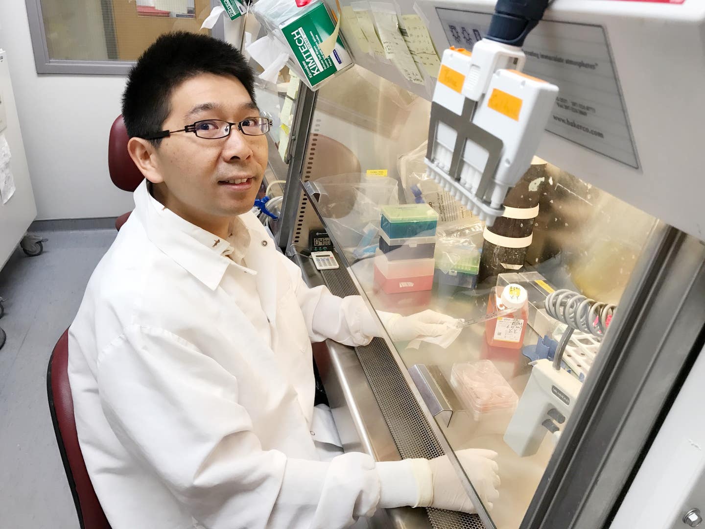
Yuanwei Yan is a scientist in the Zhang lab at UW–Madison, where researchers have developed new printing methods to grow brain tissues for use in the study of neurodevelopmental disorders like Alzheimer’s and Parkinson's diseases. (CREDIT: Xueyan Li)
A groundbreaking advancement in neuroscience has emerged from the University of Wisconsin–Madison, where scientists have developed the world's first 3D-printed brain tissue capable of growth and function like natural brain tissue.
This achievement carries significant implications for neurological research and the development of treatments for various brain disorders, including Alzheimer’s and Parkinson’s diseases.
Professor Su-Chun Zhang, from UW–Madison’s Waisman Center, highlighted the transformative potential of this breakthrough, stating, “This could be a hugely powerful model to help us understand how brain cells and parts of the brain communicate in humans.”
Zhang emphasized the potential impact on fields such as stem cell biology, neuroscience, and the study of neurological and psychiatric disorders.
Related Stories:
The innovative approach employed by the research team, outlined in the journal Cell Stem Cell, diverges from conventional vertical stacking in 3D printing. Instead, they opted for a horizontal arrangement, utilizing a softer "bio-ink" gel to house neurons derived from induced pluripotent stem cells.
Yuanwei Yan, a scientist in Zhang’s lab, explained, “The tissue still has enough structure to hold together but it is soft enough to allow the neurons to grow into each other and start talking to each other.”
The resulting brain tissue exhibited remarkable functionality, with cells forming intricate networks reminiscent of natural brain structures. Zhang highlighted the significance of this achievement, noting that even cells from different brain regions could communicate effectively within the printed tissue.
Su-Chun Zhang. (CREDIT: Andy Manis)
Compared to brain organoids, which lack organization and control, the 3D-printed tissue offers unparalleled precision in cell types and arrangement. Zhang emphasized the versatility of the approach, enabling researchers to investigate a wide range of neurological phenomena, from Down syndrome to neurodegenerative disorders like Alzheimer’s.
Zhang underscored the importance of studying the brain as a networked system, stating, “Our brain operates in networks. We want to print brain tissue this way because cells do not operate by themselves. They talk to each other.” This holistic approach, made possible by the printed tissue, promises a more comprehensive understanding of brain function and dysfunction.
Graphical abstract: Probing how human neural networks operate is hindered by the lack of reliable human neural tissues amenable to the dynamic functional assessment of neural circuits. (CREDIT: Cell Stem Cell)
Moreover, the accessibility of the printing technique opens doors for widespread adoption in research labs. Unlike complex bio-printing setups, the method requires standard equipment and can be studied using common microscopy and imaging techniques.
Looking ahead, the researchers aim to enhance their bio-ink and equipment for specialized applications. Yan expressed their intention to optimize the printing process for specific types of brain tissue, further expanding the utility of their groundbreaking technology.
Survival and differentiation of hPSC-derived neurons in fibrin hydrogel. Schematic diagram illustrating the growth of hPSC-derived NPCs in fibrin gel. hPSCs, human pluripotent stem cells; diff, differentiation; NPCs, neural progenitor cells; hGlut neurons, human cortical glutamatergic neurons. hNeurons, human neurons. (CREDIT: Cell Stem Cell)
With its potential to revolutionize the study and treatment of neurological conditions, this breakthrough marks a pivotal moment in the quest to unlock the mysteries of the human brain.
This study was supported in part by NIH-NINDS (NS096282, NS076352, NS086604), NICHD (HD106197, HD090256), the National Medical Research Council of Singapore (MOH-000212, MOH-000207), Ministry of Education of Singapore (MOE2018-T2-2-103), Aligning Science Across Parkinson’s (ASAP-000301), the Bleser Family Foundation, and the Busta Foundation.
Note: Materials provided by Brighter Side of News. Content may be edited for style and length.
Like these kind of feel good stories? Get the Brighter Side of News' newsletter.
Joshua Shavit
Science & Technology Writer | AI and Robotics Reporter
Joshua Shavit is a Los Angeles-based science and technology writer with a passion for exploring the breakthroughs shaping the future. As a contributor to The Brighter Side of News, he focuses on positive and transformative advancements in AI, technology, physics, engineering, robotics and space science. Joshua is currently working towards a Bachelor of Science in Business Administration at the University of California, Berkeley. He combines his academic background with a talent for storytelling, making complex scientific discoveries engaging and accessible. His work highlights the innovators behind the ideas, bringing readers closer to the people driving progress.


