Scientists temporarily turn skin transparent – eliminating the need for X-rays and CT scans
Stanford researchers have developed a groundbreaking method to make skin transparent using a common food dye, offering new, non-invasive ways to view internal organs.

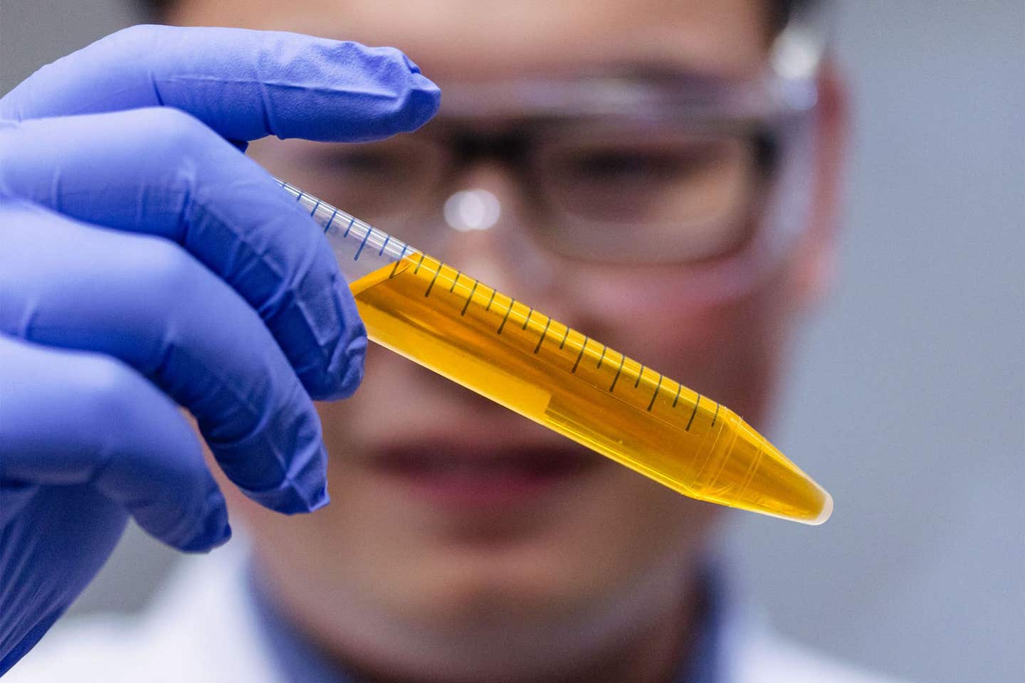
Dr. Zihao Ou, assistant professor of physics at The University of Texas at Dallas, holds a vial of the common yellow food coloring tartrazine in solution. (CREDIT: University of Texas at Dallas)
Seeing inside the human body has always been challenging. Technologies like CT scans, X-rays, and MRIs offer insights but often come with limitations. They can expose patients to radiation or fail to provide a perfectly clear picture.
But what if there was a non-invasive way to see through the skin, one that doesn’t require complex equipment or invasive procedures?
Researchers at Stanford have developed an innovative technique using an FDA-approved dye, commonly found in food, that can temporarily turn skin transparent.
Their findings, published in Science, show that by simply applying a solution of this dye onto the skin, researchers were able to peer beneath it, seeing internal organs and tissues without needing to make an incision. Just as remarkably, the effect could be reversed as easily as it appeared.
“As soon as we rinsed and massaged the skin with water, the effect was reversed within minutes,” explained Guosong Hong, an assistant professor of materials science and engineering, and senior author of the study. “It’s a stunning result.”
Related Stories:
The process works by reducing the scattering of light. Normally, when light hits the skin, it scatters, which is why skin appears opaque. This scattering happens because the refractive indices of different components in the tissue—like water and lipids—don’t match.
Water, for example, has a much lower refractive index than lipids in the visible spectrum. When light hits these mixed tissues, it bounces in different directions, making it impossible to see through them.
To counter this, the Stanford team used a solution of red tartrazine, also known as FD&C Yellow 5, a dye commonly used in food products. By massaging this dye into the abdomen, scalp, and hindlimbs of sedated mice, the researchers were able to alter how light interacted with the skin.
The dye absorbed blue light, turning the skin red. This shift in color indicated that blue light was absorbed, allowing red light to pass through more easily. As a result, the refractive index of the water in the skin began to match that of the lipids, reducing scattering and making the skin appear transparent at certain wavelengths.
The underlying physics behind this effect isn’t new. In fact, it’s based on principles described by the Kramers-Kronig relations, which link absorption and refractive index. The novelty lies in applying these principles in a biological setting, using light-absorbing molecules like FD&C Yellow 5 to change how light moves through tissue.
While this particular dye proved effective, other similar molecules produced similar results, suggesting that this method could be applied broadly.
With this technique, researchers could see internal organs like the liver, small intestine, cecum, and bladder without needing specialized imaging equipment. Blood flow in the brain and muscle fibers in the limbs also became visible.
The team even observed the beating heart and active respiratory system, demonstrating that this transparency could be achieved in living animals. Importantly, the dye did not permanently affect the skin, and once it was washed away, the transparency effect disappeared.
This non-invasive method represents a significant breakthrough in viewing living tissue without surgical procedures.
“Stanford is the perfect place for such a multifaceted project that brings together experts in materials science, neuroscience, biology, applied physics, and optics,” said Mark Brongersma, a professor of materials science and engineering and co-author of the paper. “Each discipline comes with its own language. Guosong and I enjoyed taking each other’s courses on neuroscience and nanophotonics to better appreciate all the exciting opportunities.”
While this study was conducted on animals, its potential application to humans could revolutionize healthcare. Hong explained that this approach could have significant diagnostic, biological, and even cosmetic benefits.
For example, doctors could diagnose melanoma by looking directly at skin tissue without needing to perform invasive biopsies. It could also provide a less painful method for drawing blood, as phlebotomists could locate veins more easily.
In some cases, it might even replace X-rays and CT scans, offering a clearer and safer alternative to current imaging methods. Additionally, it could improve cosmetic procedures, such as laser tattoo removal, by allowing technicians to precisely target the pigment beneath the skin.
“This could have an impact on health care and prevent people from undergoing invasive kinds of testing,” said Hong. “If we could just look at what’s going on under the skin instead of cutting into it, or using radiation to get a less-than-clear look, we could change the way we see the human body.”
In the future, this technology could provide a window into the body, allowing doctors to diagnose and treat conditions without the risks associated with more invasive procedures. While there’s still a long way to go before it’s available for human use, this research paves the way for new possibilities in medical imaging.
Note: Materials provided above by The Brighter Side of News. Content may be edited for style and length.
Like these kind of feel good stories? Get The Brighter Side of News' newsletter.
Rebecca Shavit
Science & Technology Journalist | Innovation Storyteller
Based in Los Angeles, Rebecca Shavit is a dedicated science and technology journalist who writes for The Brighter Side of News, an online publication committed to highlighting positive and transformative stories from around the world. With a passion for uncovering groundbreaking discoveries and innovations, she brings to light the scientific advancements shaping a better future. Her reporting spans a wide range of topics, from cutting-edge medical breakthroughs and artificial intelligence to green technology and space exploration. With a keen ability to translate complex concepts into engaging and accessible stories, she makes science and innovation relatable to a broad audience.


