Researchers solve the mystery of human wrinkles formation
A team of researchers has developed a method to recreate wrinkles in human tissue without animal models, offering new insights into aging, regenerative medicine, and more.
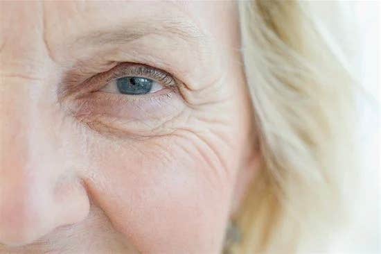
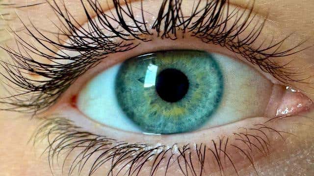
Wrinkles, though commonly associated with aging skin, are present in many organs and tissues such as the brain, stomach, and intestines. (CREDIT: CC BY-SA 3.0)
A team of researchers from POSTECH’s Department of Mechanical Engineering has achieved a significant milestone in tissue engineering by recreating the structure of wrinkles in biological tissue in vitro. Led by Professor Dong Sung Kim, alongside Professor Anna Lee and Dr. Jaeseung Youn, the research focuses on the mechanisms behind wrinkle formation.
The results were published in Nature Communications, offering new insights that could impact a range of scientific fields.
Wrinkles, though commonly associated with aging skin, are present in many organs and tissues such as the brain, stomach, and intestines. These patterns aren't merely cosmetic concerns; they play a pivotal role in regulating cellular functions and contributing to the overall physiological processes of the body.
Understanding how tissues fold and form wrinkles is essential for more than just skincare—it’s central to advancing fields like regenerative medicine and embryology.
Related Stories:
Historically, scientists have relied on animal models like fruit flies, mice, and chickens to study wrinkle formation, due to the challenges of replicating these structures in a laboratory setting. This reliance on animal models has left much of the detailed wrinkle formation process in living tissue unknown.
There has been a gap in the ability to replicate human-like tissue responses in vitro, limiting how well researchers can study and understand the formation of these wrinkle structures.
Professor Kim’s team tackled this problem by developing an epithelial tissue model entirely composed of human epithelial cells and extracellular matrix (ECM). This model was paired with a custom-built device that could apply compressive forces with precision, allowing the researchers to recreate and observe wrinkles in vitro that resemble those seen in the gut, skin, and other organs.
This model marks a breakthrough by replicating both large, deep wrinkles caused by strong compression and smaller wrinkles that form under lighter forces.
Beyond just recreating wrinkles, the team uncovered several factors that play a critical role in wrinkle formation. The porous structure of the ECM and dehydration of the tissue emerged as key contributors. By applying compressive forces to the epithelial cell layer, they observed mechanical instability in the ECM, leading to wrinkle formation.
Dehydration of the ECM layer further intensified this effect, offering an important clue into how wrinkles form, particularly in aging skin. As the underlying tissue dehydrates, it becomes more prone to developing the fine lines and deeper wrinkles associated with aging. The findings closely parallel how wrinkles form naturally, particularly in the skin as it loses moisture with age.
"We have developed a platform that can replicate various wrinkle structures in living tissue without the need for animal testing," said Professor Kim. He emphasized that this innovation allows for real-time imaging and detailed observation of how wrinkles form at both the cellular and tissue levels.
"Processes that are difficult to capture in traditional animal models can now be studied in greater detail. This has wide-ranging applications in fields such as embryology, biomedical engineering, cosmetics, and more," Kim added.
The platform not only sidesteps the ethical and logistical challenges of using animal models but also provides a more accurate representation of human tissue, making it a valuable tool for future research. It opens up possibilities for further studies in regenerative therapies, where understanding the mechanics of tissue folding could aid in developing more effective treatments for conditions that involve tissue degeneration.
Additionally, this research holds promise for the cosmetics industry, where there is a growing demand for scientific approaches to combating skin aging without relying on animal testing.
The implications extend beyond aging skin. This mechanobiological model offers a new way to study tissue folding in embryonic development and other organ systems where similar processes occur. It’s a stepping stone for advancing knowledge in biomedical engineering, potentially improving techniques for growing and repairing tissues.
The research was backed by the Mid-Career Research Program of the National Research Foundation of Korea, the Ministry of Science and ICT, and the Alchemist Project from the Ministry of Trade, Industry, and Energy.
Note: Materials provided above by The Brighter Side of News. Content may be edited for style and length.
Like these kind of feel good stories? Get The Brighter Side of News' newsletter.
Rebecca Shavit
Science & Technology Journalist | Innovation Storyteller
Based in Los Angeles, Rebecca Shavit is a dedicated science and technology journalist who writes for The Brighter Side of News, an online publication committed to highlighting positive and transformative stories from around the world. With a passion for uncovering groundbreaking discoveries and innovations, she brings to light the scientific advancements shaping a better future. Her reporting spans a wide range of topics, from cutting-edge medical breakthroughs and artificial intelligence to green technology and space exploration. With a keen ability to translate complex concepts into engaging and accessible stories, she makes science and innovation relatable to a broad audience.