Researchers make breakthrough in reversing memory loss due to head injuries
Findings suggest that the memory impairment linked to head injuries may not be a permanent affliction driven by neurodegenerative diseases

[Jan. 20, 2024: JD Shavit, The Brighter Side of News]
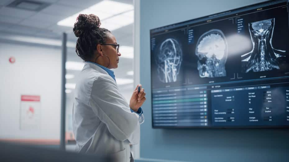
The findings suggest that the memory impairment linked to head injuries may not be a permanent affliction driven by neurodegenerative diseases. (CREDIT: Creative Commons)
In a groundbreaking study aimed at shedding light on memory loss resulting from repeated head impacts, such as those experienced by athletes, researchers have uncovered a potential breakthrough.
The findings suggest that the memory impairment linked to head injuries may not be a permanent affliction driven by neurodegenerative diseases. Instead, it could be reversible, offering hope for those suffering from cognitive impairment due to head trauma.
The study, a collaborative effort between Georgetown University Medical Center and Trinity College Dublin, Ireland, was published in the Journal of Neuroscience. It introduces a glimmer of hope for individuals who have grappled with memory deficits following head injuries.
Amnesia and poor memory after a head injury are often attributed to a lack of reactivation of neurons crucial for memory formation. However, this research indicates that the condition may not be as irreversible as previously thought.
Related Stories
Senior investigator of the study, Mark Burns, PhD, who serves as a professor and Vice-Chair in Georgetown’s Department of Neuroscience and directs the Laboratory for Brain Injury and Dementia, remarked optimistically, "Our research gives us hope that we can design treatments to return the head-impact brain to its normal condition and recover cognitive function in humans that have poor memory caused by repeated head impacts."
The researchers' previous work had uncovered the brain's adaptive response to repetitive head impacts, which altered synaptic activity and hindered memory formation. Building upon this knowledge, the team set out to investigate the possibility of reactivating forgotten memories in mice subjected to similar head impacts.
In their experiment, the scientists divided mice into two groups. One group underwent frequent mild head impacts for a week, mimicking the exposure experienced by contact sport athletes, while the other group remained unaffected as controls. Following the impacts, the mice subjected to head trauma were unable to recall a new memory a week later.
Results of tract-based spatial statistics analyses comparing the fraction anisotropy (FA) values of the patients and control groups and the correlation between post-traumatic amnesia (PTA) duration and FA values of patients. FA values were obtained for 48 regions of interest (ROIs) using a standard template of the John Hopkins University diffusion tensor imaging-based white matter atlases within the Functional Magnetic Resonance Imaging of the Brain Software Library. (CREDIT: Scientific Reports)
Burns explained the rationale behind their research focus, "Most research in this area has been in human brains with chronic traumatic encephalopathy (CTE), which is a degenerative brain disease found in people with a history of repetitive head impact. By contrast, our goal was to understand how the brain changes in response to the low-level head impacts that many young football players regularly experience."
In college football alone, players endure an average of 21 head impacts per week, with defensive ends experiencing a staggering 41 impacts weekly. The study's impact levels on mice were carefully designed to simulate a week of exposure for a college football player, with each individual head impact being exceedingly mild.
There are several initial signs that may indicate a possible serious brain injury. (CREDIT: Theraspecs)
Genetically modified mice played a pivotal role in the research, enabling the scientists to pinpoint the neurons involved in forming new memories, often referred to as "memory engrams." Surprisingly, both the control mice and the experimental mice possessed an equal presence of these memory engrams.
To delve into the physiological basis of memory alterations, Daniel P. Chapman, Ph.D., the study's first author, commented, "We are good at associating memories with places, and that's because being in a place, or seeing a photo of a place, causes a reactivation of our memory engrams. This is why we examined the engram neurons to look for the specific signature of an activated neuron. When the mice see the room where they first learned the memory, the control mice are able to activate their memory engram, but the head impact mice were not. This is what was causing the amnesia."
Ten examples of lesions causing amnesia (from total sample of 53). Lesions causing amnesia include lesions within the classic circuit of Papez and lesions outside the classic circuit of Papez. (CREDIT: Nature Communications)
Remarkably, the researchers were able to reverse the amnesia in mice by utilizing lasers to activate the engram cells. However, it's important to note that this invasive technique is not directly applicable to humans. Burns emphasized ongoing efforts to develop non-invasive methods for communicating with the brain and restoring its plasticity to potentially reverse memory loss.
This study offers a ray of hope for individuals facing memory challenges resulting from repeated head injuries, opening the door to future research and the development of innovative treatments in the quest to restore cognitive function.
Note: Materials provided above by The Brighter Side of News. Content may be edited for style and length.
Like these kind of feel good stories? Get the Brighter Side of News' newsletter.
Joshua Shavit
Science & Technology Writer | AI and Robotics Reporter
Joshua Shavit is a Los Angeles-based science and technology writer with a passion for exploring the breakthroughs shaping the future. As a contributor to The Brighter Side of News, he focuses on positive and transformative advancements in AI, technology, physics, engineering, robotics and space science. Joshua is currently working towards a Bachelor of Science in Business Administration at the University of California, Berkeley. He combines his academic background with a talent for storytelling, making complex scientific discoveries engaging and accessible. His work highlights the innovators behind the ideas, bringing readers closer to the people driving progress.


