New MRI scan can identify patients at risk for serious heart problems
UVA Health’s MRI method reveals the role of epicardial fat in heart disease, offering potential for early detection and targeted treatments.
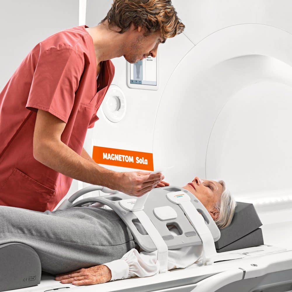
Discover how UVA Health’s innovative MRI technique uncovers hidden heart risks by analyzing the fatty composition of epicardial adipose tissue. (CREDIT: CC BY-SA 4.0)
The health risks of excess visible fat, such as that around the waist, are widely understood. However, less visible fat around the heart poses significant threats that often go unnoticed.
Scientists at UVA Health are pioneering a noninvasive way to evaluate these hidden dangers using magnetic resonance imaging (MRI). Their goal is to better understand the composition of epicardial fat and its role in heart disease.
Our hearts are naturally surrounded by a layer of fat known as epicardial adipose tissue (EAT). In healthy individuals, this fat serves as a vital protector, providing energy and insulation. However, under pathological conditions like obesity or metabolic syndrome, EAT undergoes harmful changes. It can accumulate excessively, become inflamed, and alter its fatty acid composition (FAC), contributing to cardiovascular diseases such as coronary artery disease, atrial fibrillation, and heart failure.
In a groundbreaking development, researchers led by Frederick H. Epstein, PhD, have designed an advanced MRI method to assess both the quantity and quality of this fat.
By examining the levels of saturated fatty acids (SFAs), monounsaturated fatty acids (MUFAs), and polyunsaturated fatty acids (PUFAs), they aim to predict the risks and progression of heart diseases. The potential to evaluate the proinflammatory state of EAT noninvasively represents a significant advancement in preventive cardiology.
Research, published in the journal, Magnetic Resonance in Medicine, indicates that the FAC of EAT plays a crucial role in its impact on the heart. Excessive levels of SFAs in EAT are linked to inflammation and heart disease.
SFAs activate toll-like receptor 4, triggering inflammatory responses that produce cytokines such as tumor necrosis factor alpha and interleukin-6. These inflammatory substances can damage the myocardium, leading to adverse cardiovascular remodeling and dysfunction.
Conversely, certain MUFAs and PUFAs counteract SFA-induced inflammation by inhibiting these pathways. This balance highlights the importance of identifying the fatty acid composition of EAT to guide potential treatments.
Dr. Amit R. Patel, a cardiologist and imaging expert at UVA Health, emphasized, “Using this new MRI technique, we now for the very first time have the ability to know the composition of the fat that accumulates around the heart. This is important because depending on its makeup, the fat which surrounds the heart has the potential to release damaging substances directly into the heart muscle, leading to serious heart problems.”
Related Stories
The ability to detect the proinflammatory state of EAT opens the door to early interventions. According to Dr. Patel, “We hope to show that we can convert the unhealthy fat which surrounds the heart to a more healthy type of fat with either diet and exercise or through the use of medications. We believe that by doing so, we will be able to reduce some of the complications associated with heart disease.”
Developing this imaging method was not without obstacles. The heart’s constant motion and proximity to the lungs made capturing clear images difficult. However, the UVA team overcame these hurdles by creating a rapid MRI technique that works during a single breath hold. This innovation allows precise measurement of EAT’s FAC and proton density fat fraction (PDFF).
Graduate student Jack Echols played a pivotal role in refining these computational methods. Dr. Epstein commended his work, saying, “The ability to make these measurements in epicardial adipose tissue required the use of advanced computational methods that can extract the unique signature of saturated fatty acids from an overall noisy signal. Jack Echols…did outstanding work to develop these methods.”
The UVA researchers have tested the technology in both laboratory settings and a limited group of human patients. They discovered that individuals who were obese and had experienced heart attacks showed significantly higher levels of SFAs in their EAT.
This finding suggests a direct link between fatty acid composition and heart health outcomes. “Being able to see the composition of the fat that surrounds the heart will improve our understanding of heart disease and may lead to the development of new treatment strategies in the future,” noted Dr. Patel.
The implications of this research extend beyond diagnostics. By integrating FAC mapping into routine cardiac MRI exams, healthcare providers may develop new biomarkers to noninvasively assess EAT quality. Such advancements could allow for more personalized and precise treatments, improving outcomes for patients at risk of heart disease.
EAT’s unique relationship with the heart magnifies its role in cardiovascular health. Unlike other visceral fats, EAT shares an unobstructed microcirculation with the myocardium, enabling it to directly influence heart tissue. This connection underscores the importance of understanding its composition and the inflammatory processes it can trigger.
Efforts to transform EAT’s composition through lifestyle changes or medications could be game-changing. For now, the UVA team’s innovative MRI technique represents a critical step forward in preventing and managing cardiovascular diseases.
As Dr. Epstein stated, “We’re excited to partner with cardiologists like Dr. Patel to explore clinical applications of this method, and hope that this method ultimately leads to more precise treatments and better outcomes for patients with heart disease.”
Note: Materials provided above by The Brighter Side of News. Content may be edited for style and length.
Like these kind of feel good stories? Get The Brighter Side of News' newsletter.
Joshua Shavit
Science & Technology Writer | AI and Robotics Reporter
Joshua Shavit is a Los Angeles-based science and technology writer with a passion for exploring the breakthroughs shaping the future. As a contributor to The Brighter Side of News, he focuses on positive and transformative advancements in AI, technology, physics, engineering, robotics and space science. Joshua is currently working towards a Bachelor of Science in Business Administration at the University of California, Berkeley. He combines his academic background with a talent for storytelling, making complex scientific discoveries engaging and accessible. His work highlights the innovators behind the ideas, bringing readers closer to the people driving progress.