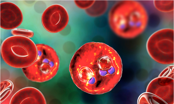
Major breakthrough in the growth, transmission and prevention of malaria
Scientists have made a major breakthrough in understanding how malaria parasites divide and transmit the disease.

[Aug 18, 2022: Charlotte Anscombe, University of Nottingham]
Researchers have made a major breakthrough in understanding how malaria parasites divide and transmit the disease. (CREDIT: Drug Target Review)
Scientists at the University of Nottingham have made a major breakthrough in understanding how malaria parasites divide and transmit the disease, which could be a major step forwards in helping to prevent one of the biggest killer infections in the world.
Malaria is still the deadliest parasitic disease worldwide, with approximately 241 million cases and over half a million deaths annually. It is caused by a single-celled parasite called Plasmodium, which is transmitted between people by the female Anopheles mosquito when they bite to take blood.
In this new study, published in PLOS Biology, scientists have uncovered the crucial roles of a group of motor proteins named kinesins during the parasite life cycle.
Different locations of kinesins in male cells of malaria parasite (Credit: The University of Nottingham)
The research, led by Rita Tewari, Professor of Parasite Cell Biology in the University’s School of Life Sciences, has shown the significance of kinesins in basic cellular processes needed for malaria parasite development, multiplication and invasion, most importantly within the mosquitoes that transmit the parasite.
Related News
Kinesins are molecular motor proteins that use energy from the hydrolysis of adenosine triphosphate (ATP - a universal store of energy in all cells), and function in various cellular processes. They are involved in transport, cell division, cell polarity and cell motility.
This latest study showed that of the nine kinesins present in the parasite genome, eight are required for the various functions of cell proliferation to cell movement in the mosquito host which was very surprising.
Researchers at the University of Nottingham have studied the location and function of all kinesins in live parasite cells at various stages of development, both in the mosquitoes which transmit the disease, and in the host where it causes disease. These proteins are important potential drug targets, hence the importance of this study in the search for new intervention targets.
(A) Life cycle of Plasmodium spp. showing different proliferative and invasive stages in host and vector. (B) Live cell imaging showing subcellular locations of representative kinesin-GFP proteins (green) during various stages of the P. berghei life cycle. DNA is stained with Hoechst dye (blue). Scale bar = 5 μm. (C) A summary of expression and location of kinesins-GFP during different stages of P. berghei life cycle. (D) Summary of phenotypes resulting from deletion of different kinesin genes at various stages of life cycle. The phenotype was examined for asexual blood stage development (schizogony), exflagellation (male gamete formation), ookinete formation, oocyst number, oocyst size, sporozoite formation in oocyst, presence of salivary gland sporozoites, and sporozoite transmission to vertebrate host. N/D, not determined.
Professor Tewari said: “This is an important genome-wide study and an essential resource for studying the various morphologically distinct parasite cells involved in parasite transmission. It shows how these important motors proteins are involved in forming molecular tracks for movement, multiplication, and transmission.”
Dr Zeeshan, who is the first author of the paper, said: “This is a comprehensive study on parasite molecular motors. It was very challenging to capture the dynamics of these proteins in live parasite cells within mosquitoes. Most importantly, we could study the formation of the male gamete (sperm), which involves a rapid multiplication process that completes within 10-12 min after the female mosquito has ingested blood from an infected host. Multiple kinesins are involved in efficient production of male gametes and deletion of kinesin genes halts parasite transmission, a discovery that can be explored further for drug discovery."
Professor Rita Tewari
“In addition, we found one motor protein, kinesin-13, which is essential for parasite multiplication in all stages of the life cycle.”
The study was carried out in collaboration with several scientists; Tony Holder at the Francis Crick Institute, London; Prof Carolyn Moores at Birbeck College; Profs Sue Vaughan and David Ferguson at Oxford Brookes; Prof Mathieu Brochet and Ravish Raspa at the University of Geneva; and Prof Karine Le Roch and Steven Abel at the University of California. This study demonstrates the power of multidisciplinary science and how networking and collaboration lead greater global understanding in science. The work was funded by BBSRC, MRC, CRUK, Wellcome Trust, NIH and NIAID.
For more science and technology stories check out our New Discoveries section at The Brighter Side of News.
Note: Materials provided above by University of Nottingham. Content may be edited for style and length.
Like these kind of feel good stories? Get the Brighter Side of News' newsletter.
Joshua Shavit
Science & Technology Writer | AI and Robotics Reporter
Joshua Shavit is a Los Angeles-based science and technology writer with a passion for exploring the breakthroughs shaping the future. As a contributor to The Brighter Side of News, he focuses on positive and transformative advancements in AI, technology, physics, engineering, robotics and space science. Joshua is currently working towards a Bachelor of Science in Business Administration at the University of California, Berkeley. He combines his academic background with a talent for storytelling, making complex scientific discoveries engaging and accessible. His work highlights the innovators behind the ideas, bringing readers closer to the people driving progress.


