Implants use smart materials to repair bone fractures
For patients with a broken shin bone, a new generation of smart orthopaedic implants is being developed that can monitor healing progression

[July 8, 2022: Claudia Ehrlich, Saarland University]
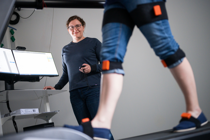
Trauma surgeon Bergita Ganse holds the Werner Siemens Foundation Endowed Professorship in Innovative Implant Development at Saarland University and is project coordinator for the 'Smart Implants' project. For patients with a broken shin bone, a new generation of smart orthopaedic implants is being developed that can not only monitor healing progression at the bone fracture site, but can use controlled micromotions to actively stimulate the repair process. At Saarland University, this innovative medical technology is being developed by an interdisciplinary team of medical specialists, engineers and computer scientists. (CREDIT: Oliver Dietze)
For patients with a broken shin bone, a new generation of smart orthopaedic implants is being developed that can monitor healing progression at the bone fracture site. And, when required, these implants will also be able to actively stimulate the repair process by undergoing controlled micromotions at the fracture site.
At Saarland University, this innovative medical technology is being developed by an interdisciplinary team of medical specialists, engineers and computer scientists. The researchers have already registered two patents and are well on the way to developing a prototype implant. The team led by Professors Bergita Ganse and Tim Pohlemann has now collated all of the available data on how mechanical stimulation can optimize the fracture healing process. The Saarbrücken team is developing implants that use shape-memory wires to generate the required micromotions. The results have been published in the journal Acta Biomaterialia.
No two fractures of the lower leg are ever the same. Whether it was a motorbike accident or a sliding tackle in football, the damage caused will differ depending on the forces that acted on the bone – and can vary from a clean break leaving two large sections of bone to a comminuted fracture where the bone shatters into multiple small pieces. And each fracture will therefore heal differently.
If you could watch a bone heal in slow motion, you would observe a fracture site that is continuously changing as new bone tissue grows. However, the typical treatment today is to screw a standard-sized orthopaedic plate to the fractured bone. These fixation plates are purely passive components. To monitor bone healing, x-ray images of the fracture site are typically taken at intervals, but this approach can only provide delayed insight into the healing process.
Related Stories
'One of the relatively common complications when using a fixation plate to mend a fractured tibia (shinbone) is that the bone does not heal properly. For every hundred patients, we typically see this problem in about fourteen cases,' said Professor Bergita Ganse. 'When using external monitoring techniques, it is difficult to identify delayed healing early enough for us to usefully intervene. This often means a prolonged route to recovery for the patient and very high costs for the health system,' explained Prof. Ganse, a trauma surgeon who holds an endowed professorship in innovative implant development from the Werner Siemens Foundation and who is coordinating the 'Smart Implants' project at Saarland University.
An interdisciplinary team of medical scientists, engineers and computer scientists are developing orthopaedic implants that can be precisely tailored to the bone of each individual patient. These innovative implants are able to supply information from the fracture site immediately after surgery and can tell the treatment team whether or not the fracture is healing properly. The implants can also issue a warning if the fracture site is subjected to incorrect loading. And, when required, this type of smart implant will be able to actively encourage the bone to heal. A prototype is planned for 2025.
The researchers are making use of the latest developments in materials technology, artificial intelligence and medical science. 'Using this new class of implants, we want to be able to continuously monitor fracture stiffness and fracture displacement at the breakage site. And if problems are identified with the healing process, the orthopaedic implant will become active and will undergo cyclic shortening or stiffening – all of which will happen without any additional surgical intervention,' explained Bergita Ganse.
Professor Bergita Ganse (left) at work in her laboratory. The qualified trauma surgeon coordinates the 'Smart Implants' project at Saarland University. For patients with a broken shin bone, a new generation of smart orthopaedic implants is being developed that can not only monitor healing progression at the bone fracture site, but can use controlled micromotions to actively stimulate the repair process. At Saarland University, this innovative medical technology is being developed by an interdisciplinary team of medical specialists, engineers and computer scientists. (CREDIT: Oliver Dietze)
In much of what they do, the researchers are working in uncharted territory. Developing an implant that is individually tailored to provide optimal support for the patient's healing process requires a thorough understanding of many complex details and relationships.
'To ensure that the bone can heal most effectively, we need to know what stimuli the smart implant needs to deliver, i.e. how much force at which frequency and in what direction will need to be applied and for how long and at what intervals,' said Bergita Ganse. That is why she and her research team have carefully collated the existing knowledge and data in the field, have described the possible mechanisms that active implants use and have identified those areas where further research is required in order to develop implants that deliver patient-specific bone repair treatment.
The results have been published in the journal Acta Biomaterialia. 'Our paper is the first to fundamentally review all of the information and data that has been published worldwide in this field,' said Ganse, who as project coordinator is also drawing on her experience as a space medicine specialist.
Ganse has contributed to research projects funded by the European Space Agency (ESA) and by the US National Aeronautics and Space Administration (NASA) that were designed to learn more about how the musculoskeletal system is affected by spaceflight. She has also helped to develop training methods for astronauts to prevent any deterioration in their bone and muscle function.
One of the most fundamental and innovative developments has been the use of shape-memory wires in orthopaedic implants. These metal fibres are able to perform tiny physical manipulations at the fracture site. But implementing this kind of technology successfully requires a huge amount of data and information. The shape-memory wires are manufactured from a nickel-titanium alloy and are no thicker than a human hair.
At Saarland University, research on these materials and their behaviour is carried out by the specialists at the Intelligent Material Systems Lab headed by Professor Stefan Seelecke. When integrated into an orthopaedic implant, these tiny electrically controlled wires can function as sensors that effectively make the healing process 'visible' or they can stimulate the bone repair process by carrying out controlled micromovements.
Shape-memory wires are able to return to their original shape after being deformed or lengthened and they can tense up and relax just like human muscle fibres. The wires are able to exert a substantial tensile force over a short distance; in fact, they have the highest energy density of all known drive mechanisms. They are powered by small electric currents. A precise electrical resistance value can be assigned to each length that these wires can assume. If the wires are integrated into an orthopaedic implant, even the smallest changes in the fracture gap can be measured.
Thanks to this capability, the wires can effectively act as sensors within the implant; with a sequence of such measurements representing movement at the fracture site. By allowing intelligent algorithms to process the large volumes of digital data generated by these sensors, motion sequences can be predicted and programmed, enabling the movement of the wires to be precisely controlled.
As a result, the implant can undergo precise changes in position at the fracture gap. It can stimulate the healing process by actively shortening or lengthening the fibres or undergoing pulsing or wavelike motions.
The research team is currently working on the fine adjustments and other important details that will enable these artificial muscles to be used in smart implants.
The research work has received €8 million in funding from the Werner Siemens Foundation.
For more science stories check out our New Innovations section at The Brighter Side of News.
Note: Materials provided above by Saarland University. Content may be edited for style and length.
Like these kind of feel good stories? Get the Brighter Side of News' newsletter.
Joseph Shavit
Head Science News Writer | Communicating Innovation & Discovery
Based in Los Angeles, Joseph Shavit is an accomplished science journalist, head science news writer and co-founder at The Brighter Side of News, where he translates cutting-edge discoveries into compelling stories for a broad audience. With a strong background spanning science, business, product management, media leadership, and entrepreneurship, Joseph brings a unique perspective to science communication. His expertise allows him to uncover the intersection of technological advancements and market potential, shedding light on how groundbreaking research evolves into transformative products and industries.


