Groundbreaking eye scanner detects Diabetes, Heart Disease, Alzheimer’s
Innovative eye-scanning technology can detect early signs of Alzheimer’s, diabetes, and heart disease through retinal scans.
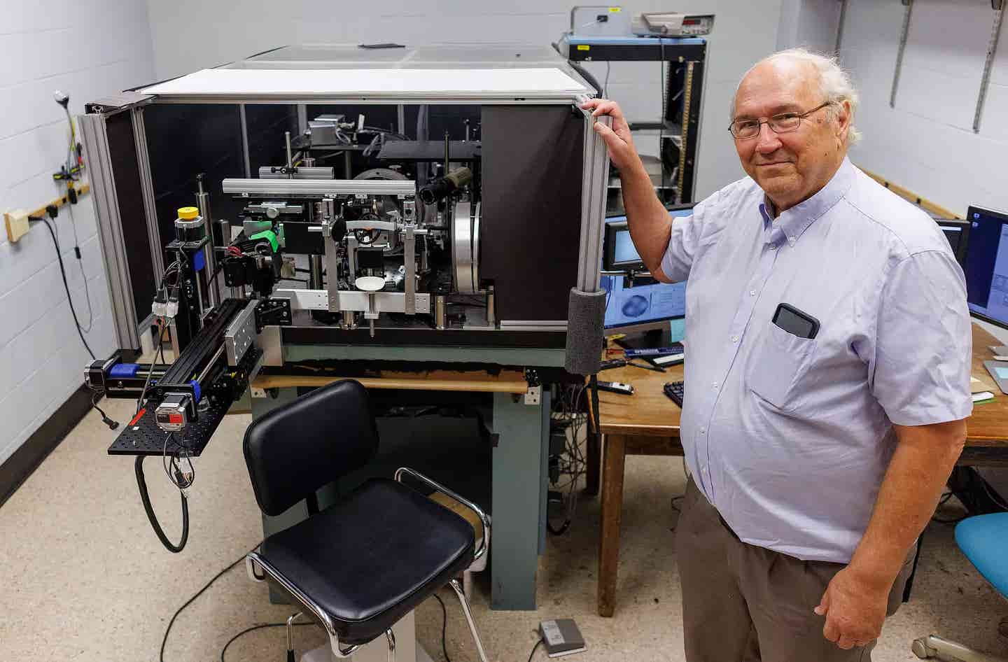
Stephen Burns stands with a high-precision ophthalmoscope capable of observing microscopic details in the back of the eye without distortion based on technology advanced at IU. Photo by Chris Meyer, Indiana University
Indiana University (IU) scientists specializing in optometry and artificial intelligence have received support from the National Institutes of Health (NIH) to develop technology in the emerging field of oculomics, which uses eye scans to detect diseases affecting the entire body.
This $4.8 million initiative, known as the NIH Venture Program Oculomics Initiative, is designed to improve diagnostic accuracy for conditions such as Alzheimer’s disease, diabetes, heart disease, kidney disease, and sickle cell anemia by examining the retina, the only visible part of the central nervous system.
Stephen A. Burns, a professor at the IU School of Optometry, leads the project along with Eleftherios Garyfallidis, an associate professor of intelligent systems engineering at IU’s Luddy School of Informatics, Computing, and Engineering.
Other key contributors include Amani Fawzi from Northwestern University, Alfredo Dubra from Stanford University, and Toco Y.P. Chui of the New York Eye and Ear Infirmary of Mount Sinai. Their work aims to develop next-generation ophthalmoscopes capable of detecting early warning signs of multiple diseases through a simple, non-invasive eye scan.
"This research is about using the eye as a window on health," said Burns, emphasizing the potential impact of this technology. The retina's visibility offers a unique opportunity to detect disease early, as the eye’s blood vessels provide a glimpse into vascular health, a key indicator of broader conditions.
Burns has been working on eye-scanning technology since the early 2000s when his team pioneered the use of adaptive optics for retinal observation. Adaptive optics, originally developed by astronomers to correct distortions in telescopic images caused by Earth's atmosphere, enable high-resolution views of the back of the eye.
Related Stories
His laboratory has refined the technology to capture real-time movements of red blood cells within retinal arteries and veins at a resolution of two microns, about one-fourth the width of a red blood cell. His team has already identified biomarkers for diabetes and hypertension in the walls of these blood vessels.
The device's potential extends beyond these conditions. For example, researchers from Northwestern and Mount Sinai have used similar techniques to observe the abnormal crescent-shaped red blood cells associated with sickle cell anemia, while Stanford’s team has focused on improving the visibility of photoreceptors in the eye. The project’s goal is to integrate these separate technologies into a single, advanced ophthalmoscope.
“There’s growing evidence of a strong retinal vascular component to Alzheimer’s disease,” Burns explained. Currently, PET scans are used to identify these signs, but they require expensive, multi-million-dollar equipment. The hope is that an eye scan could offer a far more accessible and affordable alternative.
Machine learning and artificial intelligence (AI) play a crucial role in this research. Garyfallidis, who leads the development of AI techniques for the project, is focused on training the system to analyze images rapidly and accurately, minimizing the need for human interpretation. This could reduce diagnosis time from days to mere minutes, making the technology more practical for everyday medical use.
In the first year of the project, the researchers will ensure that their instruments are calibrated to the same sensitivity levels. IU will partner with Northwestern to align their systems, while Stanford will work with the New York Eye and Ear Infirmary to do the same. This alignment is vital to ensure consistency in the data collected across different labs.
The second phase will involve validating the accuracy of the new device. Researchers will compare the new AI-driven system’s interpretations with human analyses of earlier versions of the technology. This step is essential for confirming the device’s reliability before moving on to large-scale testing.
By the final year, the team plans to test the device on clinical volunteers. Much of IU’s data will be gathered from participants recruited through the Atwater Eye Care Center. Burns highlighted the importance of this phase, noting that “up to 80 percent of the population over the age of 60 has at least one health issue that may be detectable in the eye with our technology.”
The NIH selected the project due to its potential for high impact. The NIH Venture Fund emphasizes brief, modest investments in projects with the potential to significantly accelerate scientific progress. Burns said the team’s main challenge now is refining the device’s selectivity and specificity, ensuring it can accurately distinguish between various conditions based on retinal biomarkers.
“Our challenge now is selectivity and specificity,” he said. “We need to show that we can detect the differences between conditions, to quickly and accurately interpret the signs of the various diseases we’re focusing on.”
Ultimately, the goal is to make this advanced eye-scanning technology available in everyday medical settings. Burns envisions a future where patients can undergo non-invasive scans at their annual eye exams, enabling early detection of diseases before symptoms appear.
"Wherever you get your annual eye exam, we hope this technology will eventually be available," Burns said.
By integrating machine learning with cutting-edge optical technology, this project has the potential to revolutionize medical diagnostics. Rather than relying on invasive or expensive procedures, doctors could soon detect diseases like Alzheimer’s and diabetes with a simple eye scan, giving patients a better chance at early intervention and treatment.
Note: Materials provided above by The Brighter Side of News. Content may be edited for style and length.
Like these kind of feel good stories? Get The Brighter Side of News' newsletter.
Joshua Shavit
Science & Technology Writer | AI and Robotics Reporter
Joshua Shavit is a Los Angeles-based science and technology writer with a passion for exploring the breakthroughs shaping the future. As a contributor to The Brighter Side of News, he focuses on positive and transformative advancements in AI, technology, physics, engineering, robotics and space science. Joshua is currently working towards a Bachelor of Science in Business Administration at the University of California, Berkeley. He combines his academic background with a talent for storytelling, making complex scientific discoveries engaging and accessible. His work highlights the innovators behind the ideas, bringing readers closer to the people driving progress.