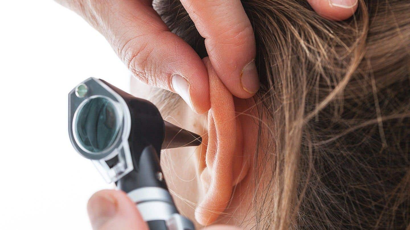
First-ever discovery of the cells in the inner ear that enable us to hear
Researchers are first to reveal that the checkerboard-like arrangement of cells in the inner ear’s organ of Corti is vital for hearing.

[Dec. 31, 2022: Verity Townsend, Kobe University]
The checkerboard-like arrangement of cells in the inner ear’s organ of Corti is vital for hearing. (CREDIT: Creative Commons)
A Japanese research group has become the first to reveal that the checkerboard-like arrangement of cells in the inner ear’s organ of Corti is vital for hearing. The discovery gives a new insight into how hearing works from the perspective of cell self-organization and will also enable various hearing loss disorders to be better understood.
The research group included Assistant Professor TOGASHI Hideru of Kobe University’s Graduate School of Medicine and Dr. KATSUNUMA Sayaka of Hyogo Prefectural Kobe Children’s Hospital.
These research results were published online in Frontiers in Cell and Developmental Biology.
Research Background
The inner ear cochlea is necessary for hearing sound, and located inside it is the organ of Corti (*1). When the organ of Corti is viewed from above under a microscope, two types of cells arranged in a precisely ordered layout resembling a chess or checkerboard can be seen. Hair cells that convey sound waves to the brain are separated by support cells, which prevent the hair cells from touching each other.
Although it has been thought that this checkerboard arrangement is necessary for the organ of Corti to function properly, the relationship between this pattern and hearing function has long remained unclear.
Related News
This research group previously revealed that this inner ear checkerboard is formed by a cellular segregation mechanism that enables the hair cells and support cells to move into line correctly. Hair cells and support cells each express a different type of the cell adhesion molecule nectin. This results in a hair cell and a support cell adhering more strongly to each other than two hair cells or two support cells would.
This property is what causes hair cells and support cells to be arranged in a checkerboard pattern. In a mouse model where one of these nectin molecules is not functional, the properties change and the checkerboard pattern cannot form correctly. In this study, the researchers used these mice to investigate the connection between the checkerboard arrangement of cells and hearing functionality.
Research Methodology
The research group compared regular (control) mice to mice with one type of nectin not functioning correctly (nectin-3 KO mouse, referred to as nectin KO mouse below). No difference between the mice was observed in the number of hair cells and support cells in the organ of Corti immediately after birth.
The organ of Corti from a normal (control) mouse. The hair cells and their support cells are lined up in an alternating, checkerboard-like pattern. (CREDIT: Frontiers in Cell and Developmental Biology)
However, there was a difference in how easily the two types of cell adhere to each other; in the nectin-3 KO mice hair cells adhere together (which does not normally happen) resulting in abnormalities in the checkerboard pattern. At this point, the researchers hypothesized that testing the hearing of these mice might reveal the relationship between hearing and the checkerboard pattern.
They measured the hearing of over one month-old nectin KO mice using the auditory brainstem response (ABR) method (*2). This test revealed that the nectin KO mice were moderately deaf, demonstrating that this hearing loss was caused by the abnormalities in the inner ear.
Statistical analysis of cell number of supporting cell, outer hair cell, and inner hair cell in nectin-3+/– or nectin-3−/− mice on P28. (CREDIT: Frontiers in Cell and Developmental Biology)
The researchers then examined the organs of Corti of the nectin KO mice that underwent the ABR test and found that the number of hair cells had decreased by approximately half. Next they set out to find out why only the hair cells (and not the support cells) had disappeared. They discovered that after 2 weeks of age, hair cell apoptosis (*3) occurred.
In addition, examination of the traces of apoptosis revealed that cell death occurred in many cells that had adhered to each other. This led the researchers to suppose that the hair cells adhering to each other (which does not normally happen) caused the apoptosis.
Progressive degeneration of outer hair cells in nectin-3 KO mice. Immunofluorescence signals for F-actin in the middle turn of auditory epithelium in nectin-3+/– or nectin-3−/− on P9, P12, and P15. Scale bars are 30 μm. (CREDIT: Frontiers in Cell and Developmental Biology)
In the epithelial tissue, which also includes the organ of Corti, there are tight junctions between each cell. These tight junctions not only connect the cells, they also prevent various molecules (including ions) from passing between the cells. If the organ of Corti doesn’t have these tight junctions, hair cells cannot function properly, cells die and hearing loss occurs.
In nectin KO mice, tight junctions were not formed properly in the places where hair cells adhered together. However, tight junctions did correctly form in between hair cells and support cells. As long as two hair cells were not adhered together, normal cell function remained. In other words, hair cell apoptosis was induced only in the places where hair cells were abnormally adhered to each other and tight junctions did not form correctly.
Apoptotic cell death of hair cells in nectin-3 KO mice. (CREDIT: Frontiers in Cell and Developmental Biology)
These results revealed for the first time that the checkerboard pattern of hair cells and support cells found in the organ of Corti functions as a fundamental structure, which protects hair cells and their functionality, by preventing hair cells from becoming attached to each other.
Further Research
Nectin is the causal gene for Margarita Island ectodermal dysplasia (*4). In addition to a cleft lip or palate and intellectual disabilities, deafness has also been reported in some cases of this genetic disorder. Therefore, the results of the current study might provide a new explanation for some cases of deafness where the cause is unclear.
This study focused on hearing and demonstrated the physiological significance of the checkerboard-like mosaic pattern of cells in the organ of Corti. However other sensory cells that respond to outside stimuli and their respective supporter cells are also arranged in the same kind of alternating mosaic pattern.
These mosaic patterns are found in sensory organs, such as the olfactory epithelium that is responsible for the sense of smell and the retina which is responsible for vision. The fact that these mosaic patterns are not only found in mammals but also in a variety of other organisms suggests that they are functionally important. The mosaic patterns in sensory tissues are created by self-organization due to the differences in adhesiveness between cells.
Therefore, focusing research on cellular self-organization in sensory organs will increase our knowledge of the functions of sensory organs and advance our understanding of various related diseases.
Main Points
In the organ of Corti in the inner ear, there are two types of cells arranged in a checkerboard-like mosaic pattern; hair cells responsible for hearing and their support cells. However, the relationship between this checkerboard pattern and hearing function has long remained unclear.
In mice in which the cells in the organ of Corti could not form into this checkerboard pattern, only the hair cells died (apoptosis), which resulted in deafness.
For the first time in the world, it was understood that the checkerboard layout plays a fundamental structural role in preserving hair cells and their functionality as the arrangement prevents hair cells from adhering to each other.
This mosaic pattern of cells has been observed in various sensory organs in many different kinds of animals. Understanding the mechanism behind how cell self-organization forms these mosaic patterns will help illuminate the functions of a variety of sensory organs and the mechanisms behind disorders.
Glossary
Organ of Corti: The sensory organ responsible for hearing. It is located inside the cochlea in the inner ear.
Auditory brainstem response (ABR): A method of recording the brain waves that are generated when sound is heard. ABR is not only used to test the hearing of newborn human babies, it can also be used on mice and other animals.
Apoptosis: A form of programmed cell death or cellular suicide that occurs in multicellular organisms.
Margarita Island ectodermal dysplasia: A genetic disorder caused by mutations in the nectin-1 gene. The main manifestation is a cleft lip or palate accompanied by intellectual disability.
Note: Materials provided above by Kobe University. Content may be edited for style and length.
Like these kind of feel good stories? Get the Brighter Side of News' newsletter.
Joseph Shavit
Head Science News Writer | Communicating Innovation & Discovery
Based in Los Angeles, Joseph Shavit is an accomplished science journalist, head science news writer and co-founder at The Brighter Side of News, where he translates cutting-edge discoveries into compelling stories for a broad audience. With a strong background spanning science, business, product management, media leadership, and entrepreneurship, Joseph brings a unique perspective to science communication. His expertise allows him to uncover the intersection of technological advancements and market potential, shedding light on how groundbreaking research evolves into transformative products and industries.


