
‘Enchanted Broomstick’ protein walks on two tiny legs to protect the brain’s nerve cells
Researchers mapped the folded structure that stops kinesin-1 from walking or carrying cargo until cells activate it.

 Edited By: Joseph Shavit

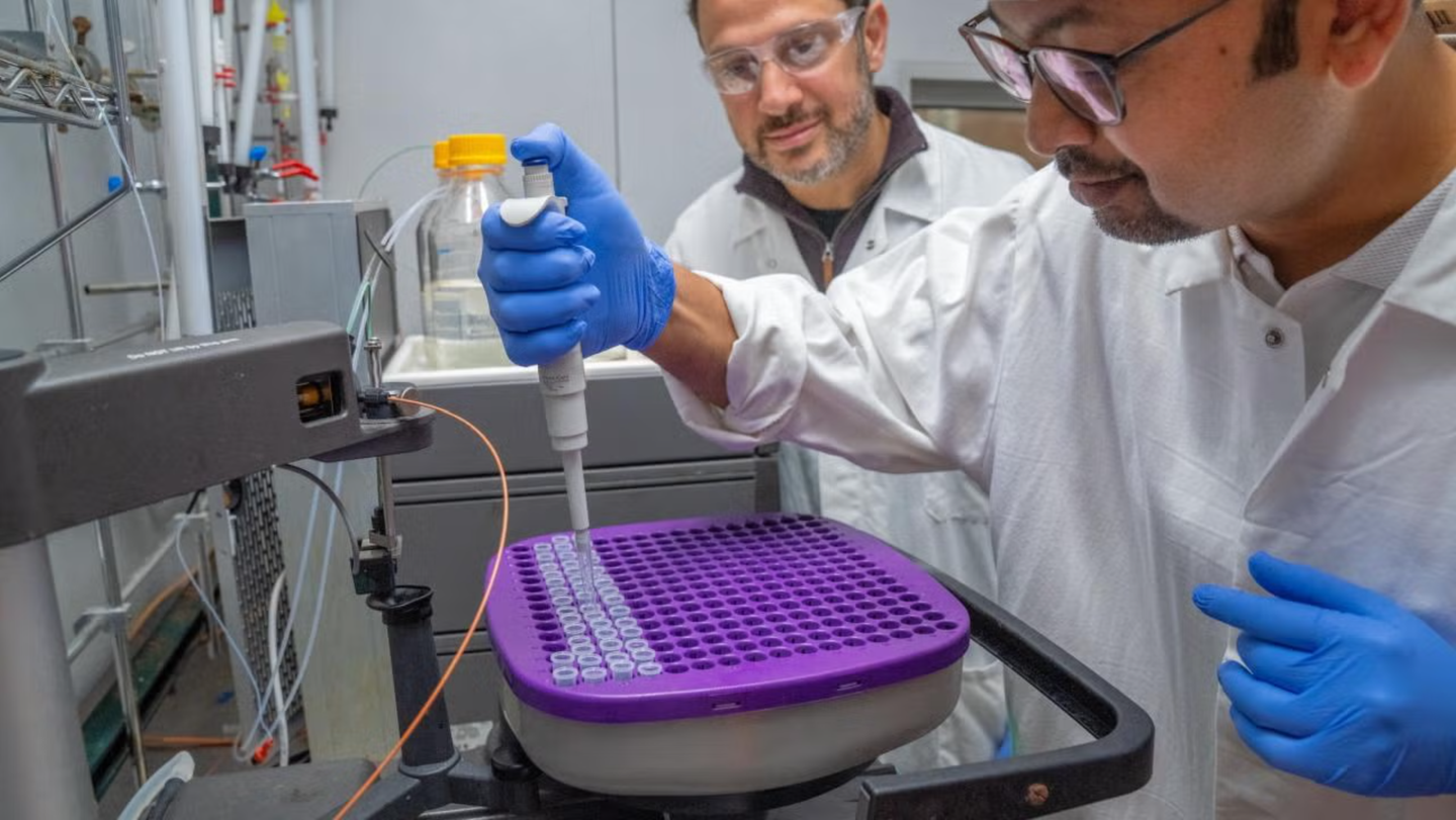
Jawdat Al-Bassam (left), associate professor of molecular and cellular biology, and postdoctoral researcher Md Ashaduzzaman helped discover how cells control a crucial “walking protein.” (CREDIT: Joaquin Benitez / UC Davis)
A nerve cell can stretch far beyond the reach of its center. Yet every distant branch still needs supplies.
Those materials travel along tiny internal tracks, carried by a protein machine called kinesin-1. It moves with two short legs and can take about 100 steps each second.
For decades, biologists understood how kinesin-1 walked. They did not fully understand how cells kept it from walking at the wrong time.
A study from the University of California, Davis, now offers a detailed answer. The work describes a folded structure that locks kinesin-1’s legs and blocks its cargo site. They could guide research into several disorders linked to faulty cell transport.
“This is the pinnacle of understanding how cells can turn on motors, precisely, to go to different places at different times,” said lead author Jawdat Al-Bassam.
Al-Bassam is an associate professor of molecular and cellular biology at UC Davis.
A Tiny Machine With a Large Job
Kinesin-1 moves along protein tracks called microtubules. These tracks extend through cells like long-distance highways.
The motor carries urgent materials away from the cell’s center. Its cargo can include neurotransmitters and other packages needed at distant sites.
This job becomes especially important in nerve cells. Their long branches can communicate with thousands of other cells. A failure in that delivery system can have serious effects. Brain cells may stop receiving the materials needed to function and survive.
Cells can then become sick or die. Such failures contribute to diseases involving paralysis, seizures, cognitive problems, blindness, weakness, and pain.
Mutations involving kinesin-1 have links to amyotrophic lateral sclerosis, Charcot-Marie-Tooth Disease Type 2, and hereditary spastic paraplegia 10. Scientists first identified kinesin-1 during the 1980s. Its unusual shape quickly caught their attention.
The protein is tall and thin, with short legs beneath a long body. Researchers have compared it to the enchanted broomstick in The Sorcerer’s Apprentice.
Its movement appears almost mechanical. Each step uses energy from a molecule called ATP. The motor breaks that molecule and uses the released energy to move forward.
“It’s a simple machine that will move in one direction if you turn it on,” said coauthor Richard McKenney.
McKenney is a UC Davis professor of molecular and cellular biology.
The problem is that kinesin-1 cannot decide when movement is useful. The cell must control every trip.
Forty Years Around One Missing Switch
Researchers already knew that kinesin-1 usually remains inactive. They also knew that a protein called MAP7 helps activate it.
MAP7 attaches to the motor and helps prepare it for movement. The physical details behind that switch remained unclear.
Al-Bassam’s group focused on the motor in its inactive form. Md Ashaduzzaman, a postdoctoral fellow, led much of the structural work. The team used cryo-electron microscopy, often called cryo-EM. This method captures many images of molecules held at very low temperatures.
Researchers gathered thousands of pictures of inactive kinesin-1. They then combined those images into a clearer structural view.
“It’s a very surprising structure,” Al-Bassam said.
The protein did not simply rest in a relaxed position. Instead, kinesin-1 folded sharply in half. Its upper end became wedged between its two motor legs.
That placement physically prevented normal walking.
A connector between the folded sections acted like a rubber band. It held the protein in its bent shape.
The design created more than one barrier. The folded upper end blocked movement near the legs. At the same time, the compact shape hid the area used for attaching cargo.
A motor without cargo would accomplish little. A loaded motor moving at the wrong time could cause serious problems. The inactive shape prevents both outcomes.
Two Locks On One Transport System
The new structure describes kinesin-1 as a tightly controlled machine rather than a freely swinging protein.
Its two legs must coordinate their motion along a microtubule. That requires enough space for their stepping cycle.
Folding the protein interferes with that motion. The motor’s cargo region also becomes unavailable. Packages cannot attach properly while the protein remains closed.
This gives the cell a double lock. One lock restrains the legs. The other blocks the loading platform.
Such control helps explain how cells manage traffic across large and crowded interiors. Cargo must leave at the correct moment and travel toward the correct destination.
The researchers then examined how MAP7 breaks this locked state.
When MAP7 attaches to kinesin-1, it wedges into the folded structure. That action loosens the connector holding both halves together.
The protein then unfolds. Its legs gain room to move. The upper cargo section becomes exposed and available for loading.
The result resembles opening a compact tool before using it. The same object changes from a locked package into an active transport machine.
Al-Bassam described the process as precise control over where and when motors operate.
That control matters because a nerve cell may need different cargo at separate branch tips. Each delivery must occur without disrupting other routes.
Disease Mutations Can Jam the Mechanism
The structural picture gives researchers a clearer way to examine disease-linked mutations.
Some mutations prevent kinesin-1 from switching on. Others may keep it active when the cell needs it to stop.
Either problem can disrupt transport. A permanently folded motor cannot deliver cargo. A motor that cannot shut down may move at the wrong time or place.
Until now, scientists lacked a detailed view of the complete inactive structure. That made it harder to see how a specific mutation could damage regulation.
The new model offers a physical map. Researchers can examine whether a mutation weakens the connector, blocks MAP7, disrupts folding, or changes the cargo site.
They can also study whether the defect comes from poor movement, poor cargo loading, or both.
“If you want to design drugs, having these clear structures will be a major advance for that,” McKenney said. “This will open up a lot of new scientific questions.”
The work does not provide a treatment. It gives researchers a foundation for asking more exact questions about diseases that currently lack cures.
“It might be possible to design a molecule that would bind the mutant protein and correct its defect,” Al-Bassam said.
Such a molecule might stabilize a motor that opens too easily. Another might help release a motor trapped in its inactive form. Those possibilities remain future goals.
A Common Design Across Living Things
Kinesin-related proteins occur throughout many forms of life. Animals, plants, fungi, and single-celled protists all use related transport systems.
That means the findings may extend beyond nerve cells or human disease.
The structure could help researchers compare how different organisms control cargo movement. It may also clarify how related motor proteins evolved.
The study included Kyoko Okada from the UC Davis Department of Molecular and Cellular Biology.
Yuqi Tang and Stephen Fried of Johns Hopkins University also contributed.
The research used the Biological Electron Microscopy Campus Core and other scientific facilities at UC Davis. The National Institutes of Health funded the work.
Practical Implications of the Research
The clearest near-term benefit is a better map of kinesin-1 regulation.
Researchers studying neurological disease can now connect particular mutations with exact parts of the motor’s locking system.
That may help separate diseases caused by poor activation from those caused by failed shutdown. Drug developers could also use the structure to search for molecules that restore normal folding or unfolding.
The work may improve broader studies of cell transport, especially inside long nerve cells that depend on precise delivery.
It also offers a useful model for understanding other molecular motors. Many cell systems rely on proteins that must remain inactive until the correct signal arrives.
For patients, any treatment remains distant. Still, drug design usually begins with a clear picture of the target.
This study supplies that picture.
Research findings are available online in the journal bioRxiv.
The original story "‘Enchanted Broomstick’ protein walks on two tiny legs to protect the brain’s nerve cells" is published in The Brighter Side of News.
Related Stories
- Weight loss connected to nerve cells in the brain, study finds
- Human touch stimulates 16 different types of nerve cells in the body
- Groundbreaking treatment repairs damaged nerve cells after injury and in chronic disease
Like these kind of feel good stories? Get The Brighter Side of News' newsletter.
Rebecca Shavit
Writer
Based in Los Angeles, Rebecca Shavit is a dedicated science and technology journalist who writes for The Brighter Side of News, an online publication committed to highlighting positive and transformative stories from around the world. Having published articles on MSN, AOL News, and Yahoo News, Rebecca's reporting spans a wide range of topics, from cutting-edge medical breakthroughs to historical discoveries and innovations. With a keen ability to translate complex concepts into engaging and accessible stories, she makes science and innovation relatable to a broad audience.