Cutting-edge AI platform significantly improves lung cancer diagnosis
Researchers develop AI platform to enhance lung cancer diagnosis and treatment, offering faster and more accurate results through digital pathology.

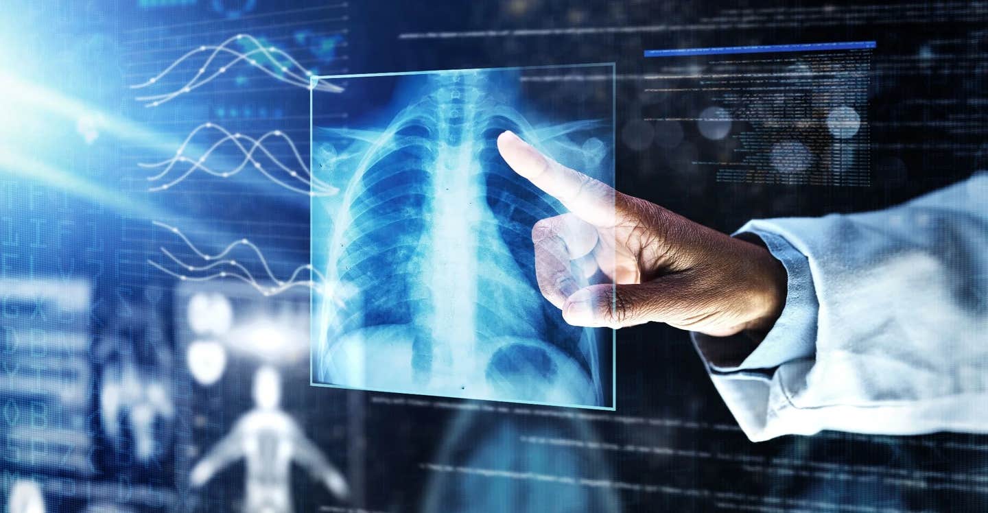
Team of researchers has made significant strides in digital pathology, particularly for lung cancer analysis.(CREDIT: PeopleImages.com – Yuri A / Shutterstock.com)
A team of researchers from the University of Cologne’s Faculty of Medicine and University Hospital Cologne has made significant strides in digital pathology, particularly for lung cancer analysis. Led by Dr. Yuri Tolkach and Professor Dr. Reinhard Büttner, the team has developed an innovative artificial intelligence (AI)-based platform.
This platform allows fully automated analysis of tissue sections from lung cancer patients, improving both the speed and accuracy of diagnostic assessments.
Lung cancer remains one of the deadliest cancers, with non-small cell lung cancer (NSCLC) accounting for more than 80% of all cases. The disease affects millions of people globally, with a particularly high incidence of lung adenocarcinoma (LUAD) in female patients, while lung squamous cell carcinoma (LUSC) is more prevalent in males.
For years, pathologists have played a central role in diagnosing lung cancer by examining tissue samples, often determining the best course of treatment based on their findings. Recently, digital pathology has transformed this process, allowing tissue sections to be digitized and analyzed on computer screens instead of under a microscope. This shift is critical for integrating advanced AI tools that can extract new types of information from these tissue samples.
Dr. Tolkach emphasized the importance of this technological leap: "The new tools can not only improve the quality of diagnosis but also provide new types of information about the patient’s disease, such as how the patient is responding to treatment."
The AI platform does more than streamline the diagnostic process; it offers insights into tumor behavior that would be impossible to detect through traditional methods. This enhanced understanding can be crucial in personalized cancer treatment, potentially altering the course of therapy based on specific molecular changes in a patient's tumor.
Related Stories
One of the major challenges in AI-based pathology, however, is ensuring that these tools meet the standards of clinical validation. While AI has been shown to be as accurate as human pathologists for several types of cancer, including prostate cancer, lung cancer remains a unique challenge.
Most AI studies for lung cancer rely on classification models, which can differentiate between tumor and non-tumor tissue but often fail to provide the detailed prognostic and predictive information needed to optimize treatment plans.
The research team behind the AI platform aims to tackle this issue by conducting a validation study involving five pathological institutes in Germany, Austria, and Japan. This will help confirm the platform’s effectiveness across different populations and environments, increasing the likelihood that it can be adopted on a global scale. Despite the promising advancements, some hurdles remain.
A major limitation is the difficulty in obtaining large, high-quality training datasets, which are essential for AI algorithms to learn from. Furthermore, the tools developed must be able to generalize well to new data, an issue that many current models struggle with. Without adequate clinical validation and quality control, these tools are unlikely to make it to market.
Dr. Tolkach’s team is hopeful that new developments in AI, such as foundation models and weakly supervised approaches, will improve the precision of digital pathology tools. Foundation models serve as powerful feature extractors from pathology images, helping to boost the accuracy of AI algorithms.
These developments, published in Cell Reports Medicine, are especially critical for lung cancer, where few AI tools exist to guide patient care. In fact, no H&E (hematoxylin and eosin)-based AI tools are currently available for lung cancer, a limitation that severely hampers the development of prognostic tools that could personalize care.
As the platform continues to evolve, the potential applications extend beyond just lung cancer diagnosis. The AI algorithms could be applied to a variety of other cancers and diseases, offering new insights that improve treatment outcomes for a wide range of patients.
The ability to automate tissue analysis could revolutionize pathology departments, making diagnoses faster and more accurate while reducing the workload on pathologists.
The future of digital pathology is promising, but the journey toward widespread clinical adoption of AI-based tools is far from complete. Only a small fraction of research in this field progresses to clinical use, and the challenges of quality control, data generalization, and explainability remain.
Nevertheless, the work being done by Dr. Tolkach and his team is an important step toward overcoming these obstacles and bringing the power of AI to the forefront of cancer treatment.
Note: Materials provided above by The Brighter Side of News. Content may be edited for style and length.
Like these kind of feel good stories? Get The Brighter Side of News' newsletter.


