Creature hiding in California’s Mono Lake sheds new light on life 650 million years ago
Discover how Mono Lake’s unique choanoflagellates offer insights into early animal evolution and their surprising symbiosis with bacteria.

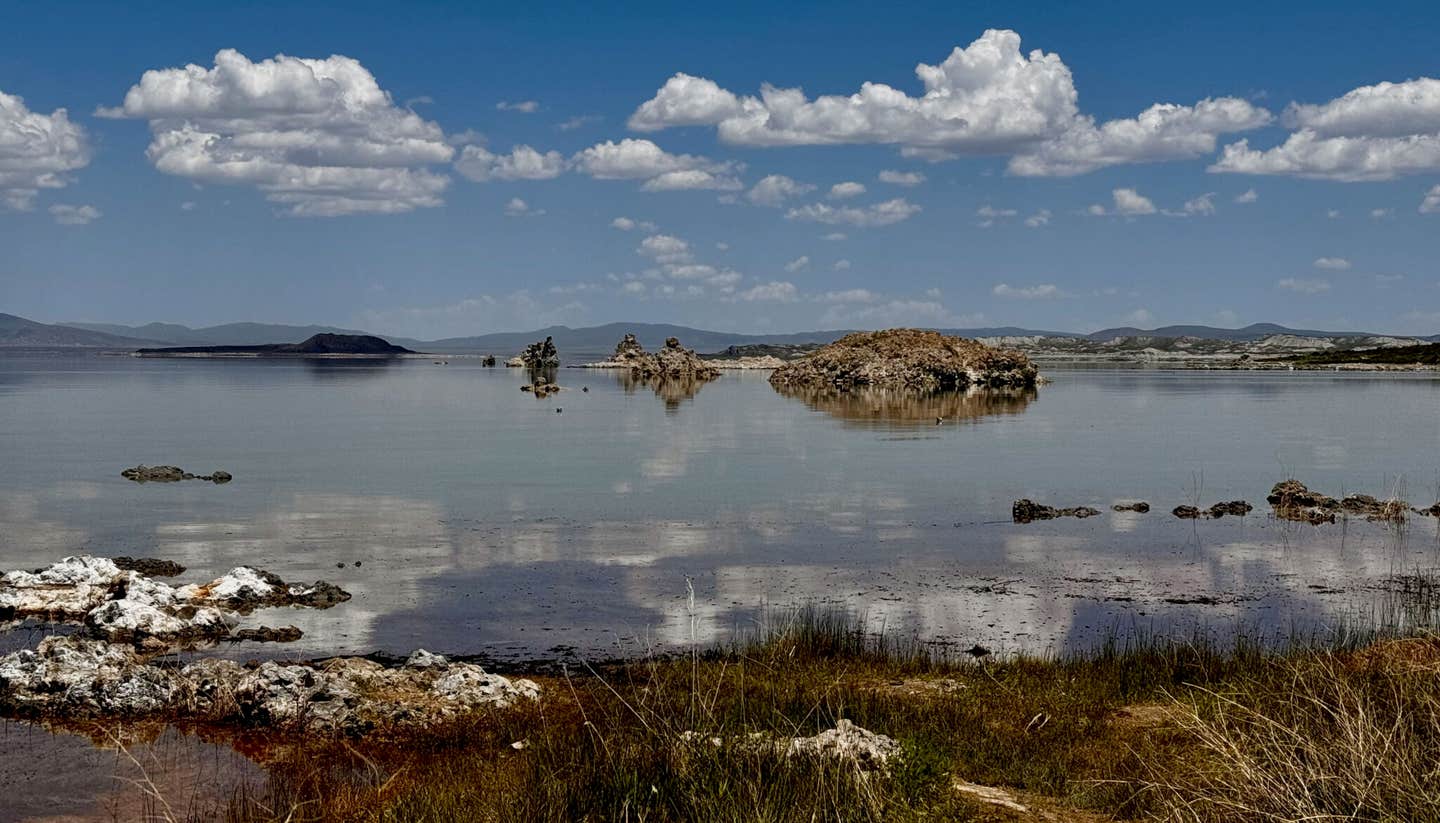
Mono Lake is located east of the Sierra Nevada, just outside Yosemite National Park. Dotted with tufa formations, the lake’s salty water is laced with arsenic and cyanide, but is home to unique flies and brine shrimp as well as choanoflagellates. (CREDIT: Nicole King)
Mono Lake in the Eastern Sierra Nevada is home to some of the most unusual lifeforms, from its towering tufa formations to the dense populations of brine shrimp and alkali flies that thrive in its harsh, salty waters.
Now, researchers from the University of California, Berkeley, have uncovered a microscopic organism within these waters that could shed light on the early evolution of life—particularly the development of multicellular organisms.
This discovery, a single-celled organism known as a choanoflagellate, has intrigued scientists. Choanoflagellates are not animals, but they are the closest known living relatives to animals, providing clues about how single-celled life transitioned into complex, multicellular organisms.
What makes this particular species remarkable is that it hosts its own microbiome—the first choanoflagellate known to form a stable, physical relationship with bacteria rather than just consuming them. As one of the simplest organisms with a microbiome, this discovery offers valuable insights into the early interactions between single-celled organisms and bacteria, which likely shaped the development of more complex life forms, including animals.
Nicole King, a professor of molecular and cell biology at UC Berkeley and an investigator for the Howard Hughes Medical Institute, explains, “Very little is known about choanoflagellates, and there are interesting biological phenomena that we can only gain insight into if we understand their ecology.”
King’s research focuses on choanoflagellates as models for early life forms that existed in ancient oceans, offering glimpses into the evolutionary history that eventually gave rise to animals.
Mono Lake’s unique environment, which includes extreme salinity and toxins like arsenic and cyanide, may have fostered the survival of this unusual choanoflagellate species. Named Barroeca monosierra by King and her colleagues, this organism could reveal how early life forms not only survived but also developed relationships with bacteria that eventually led to the complex microbiomes seen in animals today, including humans.
Related Stories
The choanoflagellate was first discovered nearly a decade ago, when Daniel Richter, then a UC Berkeley graduate student, brought back a vial of water from a climbing trip to the Eastern Sierra Nevada. Under the microscope, the water teemed with life, particularly large, beautiful colonies of choanoflagellates.
These colonies, composed of nearly 100 identical cells, formed a hollow sphere that twirled and spun as the individual cells used their flagella for movement.
King noted the similarity between these colonies and a blastula—a hollow ball of cells that forms early in the development of animal embryos. "One of the things that's interesting about them is that these colonies have a shape similar to the blastula," she said. However, it wasn’t until years later that the research team realized the full significance of the colony’s structure.
Graduate student Kayley Hake revived the choanoflagellates from a freezer and discovered something unexpected: DNA was present inside the hollow sphere, where there should have been no cells. After further investigation, she identified this DNA as bacterial, marking the first time bacteria had been observed living inside a choanoflagellate colony rather than being consumed by it.
Hake’s work uncovered not just bacteria, but also a network of extracellular matrix structures inside the colony. These structures appeared to be secreted by the choanoflagellates and could serve as a habitat for the bacteria. This was a major breakthrough because no one had ever documented a choanoflagellate forming a stable, symbiotic relationship with bacteria.
Jill Banfield, a UC Berkeley professor and pioneer in the field of metagenomics, collaborated with King’s team to identify the bacterial species found both in Mono Lake water and inside the choanoflagellate colonies. Metagenomics allows scientists to sequence all the DNA in an environmental sample and reconstruct the genomes of the organisms living there.
Banfield’s lab identified several bacterial species in the lake’s water, and Hake determined which of these were also inside the choanoflagellates. The bacterial populations inside the colonies were distinct, suggesting that some bacteria thrive better than others within the oxygen-poor interior of the colony.
While it’s still unclear whether the bacteria are being farmed by the choanoflagellates for consumption or simply taking refuge from the harsh environment of Mono Lake, King believes future research will reveal more about the interactions between these organisms. "Much of this is speculation," King admitted, but she is hopeful that the findings will provide important clues about the evolution of life on Earth.
Previous studies in King’s lab have already shown that bacteria can influence choanoflagellate behavior, including stimulating mating and encouraging the formation of colonies. Barroeca monosierra will likely serve as a new model system for studying the interactions between eukaryotes (organisms with complex cells) and bacteria, as well as the role bacteria played in early animal evolution.
Despite these promising findings, Mono Lake’s choanoflagellate colonies are elusive. During a recent visit, only six out of 100 water samples contained the choanoflagellates. Nonetheless, King remains excited about the future of this research. "I think there's a great deal more that needs to be done on the microbial life of Mono Lake because it really underpins everything else about the ecosystem," she said.
King’s work, along with that of her colleagues, could help answer fundamental questions about the relationships between early life forms and their microbial companions—relationships that likely paved the way for the human microbiome and the complex interactions between animals and bacteria that we see today.
In addition to King and Banfield, other contributors to the research include graduate student Kayley Hake, former doctoral student Patrick West, electron microscopist Kent McDonald, and postdoctoral fellows Josean Reyes-Rivera and Alain Garcia De Las Bayonas. Their work is supported by the Howard Hughes Medical Institute and the National Science Foundation.
Note: Materials provided above by The Brighter Side of News. Content may be edited for style and length.
Like these kind of feel good stories? Get The Brighter Side of News' newsletter.
Joshua Shavit
Science & Technology Writer | AI and Robotics Reporter
Joshua Shavit is a Los Angeles-based science and technology writer with a passion for exploring the breakthroughs shaping the future. As a contributor to The Brighter Side of News, he focuses on positive and transformative advancements in AI, technology, physics, engineering, robotics and space science. Joshua is currently working towards a Bachelor of Science in Business Administration at the University of California, Berkeley. He combines his academic background with a talent for storytelling, making complex scientific discoveries engaging and accessible. His work highlights the innovators behind the ideas, bringing readers closer to the people driving progress.


