AI-powered ‘Alzheimer’s in a Dish’ model revolutionizes new drug development
Discover how innovative 3D models and algorithms are revolutionizing Alzheimer’s drug discovery, offering new hope for effective treatments.

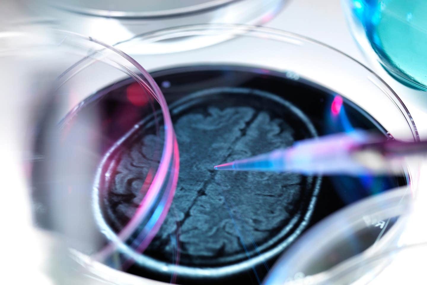
A groundbreaking combination of 3D cell culture models and computational tools is accelerating Alzheimer’s drug discovery, offering renewed hope for effective therapies. (CREDIT: ALAMY)
Alzheimer’s disease remains one of the most pressing challenges in modern medicine, ranking as a leading cause of death worldwide with no proven treatments to reduce mortality. The disease is characterized by a complex interplay of amyloid-β deposits, tau tangles, neuroinflammation, and widespread neuronal loss.
Despite decades of research, most drugs that show promise in laboratory and animal models fail during human trials, leaving researchers grappling with fundamental questions about why these failures occur.
One major issue lies in the inherent complexity of Alzheimer’s pathology, which involves multiple interconnected pathways rather than isolated molecular events. This complexity makes it difficult to identify reliable drug targets or create preclinical models that accurately replicate human disease.
Traditional models, particularly those using rodents, fail to mimic critical aspects of Alzheimer’s, such as amyloid-β-driven tau pathology, limiting their ability to predict drug efficacy in humans. As a result, researchers have turned to human-derived models and computational tools to close the gap between laboratory findings and clinical success.
A significant breakthrough came a decade ago with the development of a 3D cell culture model known as "Alzheimer’s in a dish." This system uses human neural precursor cells suspended in gel to simulate the progression of Alzheimer’s over a decade in just six weeks.
Unlike animal models, these human-derived systems reflect the genetic diversity and complexity of Alzheimer’s pathology, offering a more accurate platform for drug testing. However, doubts remained about whether these models truly replicated the molecular and functional changes observed in human brains affected by Alzheimer’s.
To address this uncertainty, a team of researchers from Mass General Brigham and Beth Israel Deaconess Medical Center developed a computational tool called integrative pathway activity analysis, or IPAA.
Their results, published in Neuron, identify crucial shared pathways, confirming that the Alzheimer’s in a dish model can be used to assess new drugs accurately and rapidly as well as point the way to drug discovery.
This platform evaluates how closely Alzheimer’s models replicate the gene expression patterns and biological pathways seen in patient brains. By focusing on broader biological pathways instead of individual genes, IPAA offers a more precise understanding of the disease's molecular landscape.
Related Stories
The researchers used this tool to compare gene expression data from deceased Alzheimer’s patients with data from 3D cellular models. Their findings identified 83 dysregulated pathways common to both human brains and the Alzheimer’s in a dish model, confirming the model’s ability to replicate key aspects of the disease.
This approach not only validated the Alzheimer’s in a dish model but also revealed actionable targets for drug development. For example, the researchers focused on the p38 mitogen-activated protein kinase (MAPK) pathway, which was significantly dysregulated in both patient brains and the 3D model.
Testing a clinical p38 MAPK inhibitor, they found that the drug effectively reduced Alzheimer’s pathology in the dish, highlighting its potential for clinical trials. This discovery underscores the power of combining human-derived models with computational tools to identify promising drug candidates more efficiently.
Dr. Doo Yeon Kim, a senior researcher from Massachusetts General Hospital and co-developer of the 3D model, emphasized the importance of this validation. “Our goal is to find the best model with the most similar activity to what we see in the brains of patients with Alzheimer’s disease,” he explained. “We developed this 3D cell culture model for Alzheimer’s 10 years ago. Now we have the data that show that this model can accelerate drug discovery.”
This breakthrough was the result of collaboration between neurologists and computational scientists, who combined their expertise to overcome longstanding barriers in Alzheimer’s research.
Dr. Winston Hide, a senior researcher at Beth Israel Deaconess Medical Center, described the transformative nature of their work. “We faced a fundamental challenge: understanding which models truly reflect the complexity of Alzheimer’s in the human brain. By shifting focus from individual genes to the broader biological pathways they shape, we’ve created a system that transforms how we discover and test drugs.”
The implications of this research extend beyond a single pathway or model. The IPAA platform enables researchers to systematically evaluate multiple drugs and pathways simultaneously, significantly speeding up the drug discovery process.
Already, hundreds of approved drugs and natural compounds have been tested using the Alzheimer’s in a dish model, paving the way for future clinical trials.
Dr. Rudolph Tanzi, another senior researcher and Director of the McCance Center for Brain Health, highlighted the potential impact of these advancements. “Now we have a system that not only allows us to test new drugs quickly but also an algorithmic platform that can predict which drugs will work best. Together, these advancements bring us closer to finding better drugs and getting them to patients.”
By combining innovative 3D models with cutting-edge computational analysis, researchers have created a powerful system for understanding Alzheimer’s disease and accelerating the search for effective treatments. This integrated approach offers renewed hope for a field that has long struggled with high failure rates and limited progress.
As the technology continues to evolve, it may finally bring much-needed breakthroughs to patients and families affected by this devastating condition.
Note: Materials provided above by The Brighter Side of News. Content may be edited for style and length.
Like these kind of feel good stories? Get The Brighter Side of News' newsletter.
Joshua Shavit
Science & Technology Writer | AI and Robotics Reporter
Joshua Shavit is a Los Angeles-based science and technology writer with a passion for exploring the breakthroughs shaping the future. As a contributor to The Brighter Side of News, he focuses on positive and transformative advancements in AI, technology, physics, engineering, robotics and space science. Joshua is currently working towards a Bachelor of Science in Business Administration at the University of California, Berkeley. He combines his academic background with a talent for storytelling, making complex scientific discoveries engaging and accessible. His work highlights the innovators behind the ideas, bringing readers closer to the people driving progress.


